 | Diagnosing a Meniscus InjuryMedical professionals (such as an orthopedic surgeon or physician) will be able to test whether you have a meniscus injury and then will determine what type you have through a variety of assessments. It is more difficult to diagnose a lateral meniscus tear than a medial meniscus tear because of its tear shape and location (it may go unnoticed until it is much larger). To help your doctor achieve a proper diagnosis, he/she will begin with a medical history about you, your current condition and symptoms, the intensity of your pain, the duration of your symptoms and the limitations you are experiencing. Details about what instigated the problem, when it started, and whether or not you have ever had treatments (for this or a similar condition in the past) are very helpful in assessing your injury. Common Meniscus Physical Examinations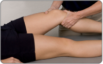 A physical examination will be performed to determine if you have any signs of a meniscus injury or possibly another knee injury. Your doctor will visually assess and palpate (feel) the bones and soft tissue in and around both of your knees to evaluate symmetry and spot any differences. This will identify abnormalities such as inflammation, bone deformity, and atrophied muscles. He/she will press on the injured side of your knee joint to test for point tenderness and help determine the location of your injury or tear. He/she may ask you to complete a series of knee and leg movements such as moving your knee from a straight to bent position (or vice versa), or rotating your knee to see what motions cause pain, weakness, instability and/or grinding, catching, popping or locking. Your knee will also be inspected for fluid, swelling and warmth. McMurray's Test is performed while you lie flat on your back. The doctor will hold your knee with one hand and your ankle in the other. He/she will lift your knee slightly while flexing it to a 45 degree angle. The doctor will feel the medial joint line while pulling your leg toward him/her and rotating your knee. The knee is then brought from full flexion to 90 degrees. If you feel a click during this movement the test is positive; you have a meniscus injury. Joint Line Tenderness is simply performed by applying pressure over the meniscus area while you are laying flat on your back. If pain occurs, the test is considered positive. Ege's Test is performed while you are standing and putting weight on your knees. You begin by standing with your feet 8-10 inches apart. To test the lateral meniscus the doctor will ask you to turn your feet and knees in as far as possible; for medial meniscus tests, you turn your feet and legs out fully. In the appropriate position, you will then squat down and stand up slowly. If you experience pain and/or a clicking sensation (you may even hear it) when your knees are at approximately 90 degrees, the test is considered positive and a meniscus tear is suspected. Common Meniscus Tear Diagnostic Tests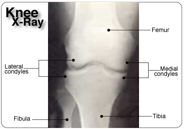 A medical professional will sometimes recommend diagnostic testing to obtain more detailed information, and assess the amount and/or type of damage done to your knee and meniscus. There are a variety of different tests available to help them analyze the situation; however the recommendation will be dependent on your injury. X-rays will provide an image of the overall structure of your knee. It is helpful in identifying abnormal bone shapes, fractures, arthritis, and degeneration (wear and tear) within the joint. It can identify a discoid meniscus, or loose bones and bone abnormalities that may mimic a torn meniscus. CAT scans (or CT - computerized tomography) can be used to provide a 3-dimensional assessment of the bones and soft tissues in and around your knee joint and may be used to identify a meniscus tear.
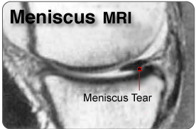 MRIs (magnetic resonance imaging) will provide more detailed information and will help to evaluate the soft tissues in and around your knee joint (muscles, tendons, ligaments, menisci, other connective tissues). It can identify ligament and meniscal damage, and help to determine the extent of your injury, the displacement and degree of your tear, fluid on your knee, a discoid meniscus, ACL or MCL tear, and/or other associated conditions. The type of test recommended will depend on your symptoms and the opinion of your medical professional. Further tests such as diagnostic ultrasound, electromyogram, or arthroscopic surgery can be used to determine the degree and location of your injury if required. Types of Meniscal TearsThe severity of your meniscus tear will vary depending on which meniscus is injured and the location, type and shape of the tear. Tear LocationBlood supply to the injured area is critical to healing; where the tear is located will determine its ability to heal. The fibrocartilage of the meniscus has limited blood flow compared to other soft tissue in the body and this can make it difficult for the body to heal a meniscus tear on its own. The amount of blood vessels throughout the meniscus varies. The meniscus can be broken into the red zone (outer portion of the meniscus that is vascular), the middle (central part with fewer blood vessels), and the white zone (inner third containing no blood vessels); tears in the red zone have the best chance of healing because they have more access to blood supply. 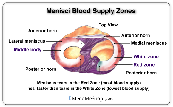
Tear TypeYour knee joint can also be divided into the anterior horn (mobile, curved portion at front of meniscus), the posterior horn (less mobile, curved portion at back of the meniscus) and the body (middle section of the meniscus, thicker on outside and thinner on the inside). 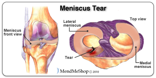 There are 3 types of meniscal tears you can experience in these locations:
Tear ShapesThe shape of your meniscus tear is important because the tear shape will help determine the type of treatment you receive; some tears will heal without surgery, some can be treated surgically and some can't be fixed. Tears come in many shapes and sizes, however there are 3 basic shapes for all meniscal tears: longitudinal, horizontal, and radial. If these tears are not treated, they may become more damaged and develop a displaced tear often referred to as a bucket handle tear (longitudinal), flap tear (horizontal) or parrot beak tear (radial). Complex tears are a combination of two or more of these basic shapes with damage occurring in more than one direction and depth. Longitudinal Tear (Circumferential Tear)A longitudinal tear extends lengthwise, following the collagen fibers that run parallel to the contour of the meniscus. This tear does not go all the way through the meniscus and it divides your meniscus into an inner and outer section; however the tear generally never touches the rim of the meniscus. It tends to be more medial than lateral and results from repeated movements. It generally starts as a partial tear in the posterior horn, which can sometimes heal on its own. 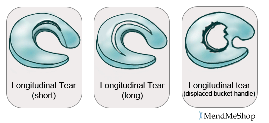 If a longitudinal tear doesn't heal properly it can lead to a displaced tear, known as a bucket handle tear. This is a complete tear that goes all the way through and never touches the inner rim of your meniscus. There is a risk that the handle may flip over and can catch on the femur, locking the joint and increasing pain. This tear accounts for 10% of all meniscus tears, and causes your knee to lock in flexion. It is seen most often in young athletes, and happens in conjunction with 50% of ACL injuries. Horizontal Tear (Cleavage Tear)A horizontal tear starts as a horizontal split deep in your meniscus. This tear divides your meniscus into a top and bottom section (like a sliced bun). It is often not visible and moves from the posterior horn or mid section to the inside of your meniscus. Horizontal tears are rare and often start after a minor injury from rotation or degeneration. It occurs most often in your lateral meniscus but however it is noted in both menisci. 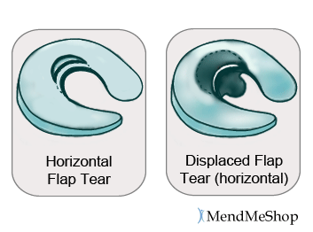 A displaced horizontal flap tear can develop if your horizontal tear is overlooked or not cared for. This type of tear is horizontal on the surface of your meniscus and creates a flap that flicks when your knee moves. It is a result of a strong force that tears your meniscus from the inner rim; it can easily become a complex tear if left untreated. Often, the flap is trimmed away during surgery to prevent further tearing. Since the periphery of the meniscus is not compromised and there is enough tissue left to heal, the cushioning function of the meniscus is maintained. If this tear extends from the apex of your meniscus to the outer rim, you may develop a meniscal cyst (a mass that develops from a collection of synovial fluid along the outside rim of the meniscus). Radial Tear (Free-Edge Transverse Tear)A radial tear starts as a sharp split along the inner edge of your meniscus and eventually runs part way or all the way through your meniscus, dividing it into a front and back section (across the middle body instead of down the length). This tear generally occurs between the posterior horn and middle section and is seen frequently in your lateral meniscus. 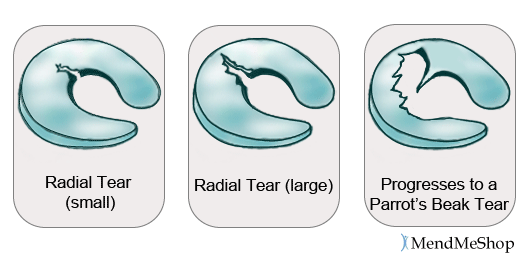 A small tear is difficult to notice, but when it grows and becomes a complete tear it will open up and look like a part is missing. This is called a Parrot's Beak tear (displaced radial tear with a curved inner portion). It generally occurs in the thicker portion of your lateral meniscus. As it gets larger, it will catch or lock more frequently, and prevent your meniscus from protecting the articular cartilage during weight bearing. This tear is the result of a traumatic event or forceful and repetitive stress activities and it is often associated with other injuries such as ACL tears. Young athletes tend to suffer from combination tears called radial/parrot beak tears (the meniscus splits in 2 directions). To learn about conservative home treatments options for meniscus injuries or other soft tissue knee injuries visit our meniscus treatment page.
Product Advisors are available 9:00 am to 5:00 pm Eastern Standard Time Monday to Friday. Learn More About Knee Injuries & TreatmentsI want to learn more about post-surgery recovery. I want to learn all about common causes of knee pain. I want to learn more about TShellz Wrap® Circulatory Boost I want to learn more about Ice & Heat: Which Is Better For Treatment? I want to learn more about Knee Treatments. I want to learn more about Runners Knee. I want to learn more about Hoffas Syndrome. I want to learn more about Knee Arthritis. I want to learn more about Knee Trigger Points. FREE SHIPPING ON ALL PRODUCTS CURRENTLY ENABLED |
    |